3dMD, which uses an array of highly sensitive cameras and can image the entire body in a fraction of a second, generates extremely accurate and reliable models of the torso that can guide the management of adolescents with scoliosis, according to a new study that includes researchers at Hospital for Special Surgery (HSS) in New York City.
The findings, presented at the American Academy of Orthopaedic Surgeons (AAOS) 2022 Annual Meeting, suggests that the ultrafast cameras produce scans of the surface of the body that complement internal images generated by low-dose x-ray. Because 3dMD does not rely on ionizing radiation, the investigators believe the technology may help scoliosis patients avoid many of the repeated and potentially harmful x-rays that conventional care require.
Surface topography and internal imaging work together to provide a much fuller picture of how scoliosis affects the body, according to Roger F. Widmann, MD, chief of the Pediatric Orthopedic Surgery Service at HSS and the senior researcher on the study. “When you see someone with a spinal deformity, you’re not necessarily visualizing the shape of the spine underneath,” he says. “Three-dimensional (3-D) surface scanning is analogous to what you see visually, whereas x-rays or MRI are often much different from what you see on the surface. Both approaches are important, but the information they provide is quite different.”
Howard J. Hillstrom, PhD, a biomechanical engineer at HSS and co-principal investigator on the project, says the 30-camera array can generate a full image of a standing person in under two milliseconds—fast enough to eliminate any blurring from even the slightest movement in any direction. “It gives us very accurate information to begin with, and it’s important to start with sound data,” Dr. Hillstrom notes.
What was previously unknown was how well these images of the body’s surface conform to the anatomy beneath the skin. This relationship is critical for 3dMD to be useful in the clinic as a way to guide treatment for scoliosis, which affects between 2% and 3% of youth in the United States, or 6 to 9 million people.
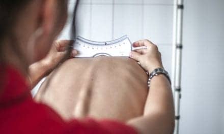
Treatment for scoliosis frequently involves the use of braces to correct the curvature of the spine—a measurement called the Cobb angle—but in the most severe cases, surgery may be necessary.
Dr. Hillstrom, who describes the image as a “large, 3D selfie,” said these results “give us the green flag to study spinal pathology and the effects of treatments for different severities of scoliosis” in ways that weren’t possible previously.
For clinicians, he adds, “It’s great to have objective tools to support the patient-reported outcomes that we use.”
3dMD vs EOS
For the study, the researchers compared images gathered by 3dMD with those produced by a next-generation x-ray system called EOS. A total of 46 youths participated in the study, 26 of whom had been diagnosed with adolescent idiopathic scoliosis (mean age, 14.7 years; 14 female, 12 male) and 20 who did not have the condition (mean age, 14.6 years; 9 female, 11 male).
The 3dMD images demonstrated extremely high reliability, particularly for linear geometric measurements of the surface of the subjects. “The measurements generated from the 3-D scans were used to create unique volumetric measures of truncal asymmetry, which demonstrated very high correlations with patient-reported self-image,” Dr. Widmann said.
Dr. Hillstrom notes the post-processing software that employs artificial intelligence (AI) is the secret behind the accuracy of the camera array. “AI fills in the blanks very quickly. It helps to identify anatomical regions and provide a mathematical approach to organizing the topographic data showing you the head, neck, trunk and pelvis—from outside of the body. You can uncover details you wouldn’t normally be able to see with the naked eye or with the use of conventional radiology.”
As the 3dMD system moves into the clinic, the technology will help surgeons better predict both the cosmetic and functional outcomes of the various procedures for scoliosis. “You can use the system to assess the efficacy of different surgical techniques based on how well you’re creating symmetry of the surface of the torso,” Dr. Widmann says. “This technology offers a quick and easy way to objectively assess surface symmetry as well as global spine range of motion, and both measures are really important to assess after scoliosis surgery.”
[Source(s): Hospital for Special Surgery, PR Newswire]
Related Content:
Drug Combo Reduces Narcotic Use, Manages Pain After Scoliosis Surgery
For Kids with Severe Scoliosis, ‘One and Done’ Surgery Has Advantages, Study Notes