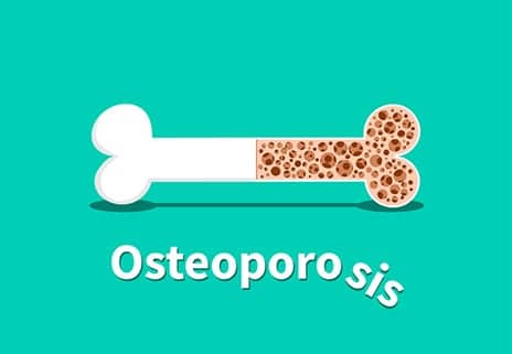
Researchers from University of Pennsylvania’s School of Veterinary Medicine (Penn Vet) suggest a mechanism by which certain risk factors, such as smoking cigarettes, consuming alcohol, taking certain medications, or being exposed to environmental pollutants, could lead to the development of osteoporosis.
Damage to mitochondria—key cellular organelles and energy generators—leads to a surge in the creation of cells called osteoclasts, which are responsible for breaking down bone, the researchers report in a study published recently in The FASEB Journal. They uncovered these effects in cells in culture as well as in an animal model.
“In a normal individual, the process of bone degradation and rebuilding proceeds in a very balanced way, but in some people they somehow produce a lot more osteoclasts, and this leads to bone loss and osteoporosis,” says Narayan Avadhani, a biochemist at Penn Vet and senior author on the work.
“We show in this paper that, when mitochondrial function is affected, it not only affects energy production but also triggers a type of stress signaling that induces the overproduction of osteoclasts,” Avadhani adds, in a media release from University of Pennsylvania.
In collaboration with the lab of Mone Zaidi at Icahn School of Medicine at Mount Sinai, the scientists in Avadhani’s lab took a close look at how problems with mitochondria affected a type of immune cell known as macrophages.
To understand how mitochondrial damage could be linked to osteoporosis through the work of macrophages, the researchers induced damage to a key enzyme responsible for energy production in mitochondria, cytochrome oxidase C, in lab-grown mouse macrophages. Doing so led the macrophages to release a variety of signaling molecules associated with an inflammatory reaction and also seemed to encourage them to go down the path toward becoming osteoclasts.
Looking closely at what was going on, they observed an anomaly with a key molecule, RANK-L, that helps regulate the bone-rebuilding process and is released by bone-building cells as a means of inducing bone breakdown. When mitochondria were damaged, they underwent stress signaling and transformed into osteoclasts at a much faster rate, even when RANK-L levels were low. These osteoclasts led to greater rates of bone resorption, or breakdown, the release explains.
“In some respects, mitochondrial stress signaling may even be replacing RANK-L,” Avadhani notes. “That we don’t know now, but we plan to look into that further.”
The researchers confirmed their findings in a mouse model, showing that animals with a mutation that leads to dysfunctional mitochondria had increased production of osteoclasts. In addition, their macrophages had higher levels of phagocytosis, the process by which the immune cells engulf and “eat” invaders. The researchers believe this enhanced phagocytic ability could be responsible for some of the other physiological problems that can arise in people with defects in their mitochondria.
Because some of the same environmental risk factors that seem to promote osteoporosis, like smoking and some pharmaceuticals, can also impact mitochondrial function, the team posits that this stress signaling might be the pathway by which they are acting to affect bone health.
Future studies by the group, including Mount Sinai’s Zaidi, a specialist in various aspects of osteoclast function and bone health, may look at how safeguarding mitochondrial function could protect against osteoporosis, per the release.
[Source(s): University of Pennsylvania, EurekAlert]


