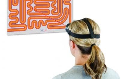
PHOTO CAPTION: A patient completes a knee kinesiography examination using the KneeKG to evaluate knee dysfunction.
Pain treatment modalities complemented by targeted exercises and movement training can provide physical therapists with a winning combination for osteoarthritis patients’ knee pain.
By Joseph A. Zeni Jr, PT, PhD
Knee osteoarthritis (OA) is one of the most prevalent orthopedic conditions in older adults. Although the primary complaint of patients with knee OA is joint pain, this condition also leads to a variety of other impairments and deficits. These include muscle weakness, altered movement patterns, decreased physical activity, and difficulty with activities of daily living. Therefore, it is important that physical therapists take a multimodal approach when caring for these clients.
Many osteoarthritis treatments target joint pain, but therapists should also address strength, range of motion, and biomechanical deficits, such as varus thrust, to have the best chance of improving patient outcomes and preventing OA progression.
Management and Modalities
Pain management for osteoarthritis typically includes physiotherapy. Therapists will prescribe exercises to improve strength and range of motion, as well as provide modalities to reduce swelling and pain. Compression knee wraps, massage, icing (cryotherapy), heating (thermotherapy), and acupressure can be used in combination with active exercise therapies to reduce pain and improve function. These modalities often offer temporary relief of acute and chronic pain.
Physical therapists have a variety of non-addicting, non-invasive approaches they may use to provide temporary pain relief. Transcutaneous electrical nerve stimulation, for example, transmits low-voltage electric current through the skin to activate endorphins that subsequently block the perception of pain. Electrical stimulation (E-Stim) devices are another type of tool that provides temporary pain relief, by sending electrical pulses through the skin that block pain signals from reaching the brain. Both TENS and E-Stim can be used to temporarily relieve arthritis pain.
Other non-invasive options include hot/cold therapy packs that provide moist heat and therapeutic cooling. Battle Creek Equipment, Freemont, Ind, provides a line of such products including Thermaphore Classic Moist Heat Packs and Ice It! Cold Therapy. Another source is Southwest Technologies, North Kansas City, Mo, which offers Elasto-Gel hot and cold therapy products, designed to provide moist therapy heat or soothing cold.
Another option for short-term relief are topical analgesics formulated in gels, creams, and sprays. Topical analgesics provide an affordable treatment solution to penetrate the skin and modulate an individual’s perceived level of pain. Several sources offer topicals to the PT market, including Sore No More, Moab, Utah, which offers Sore No More Natural Pain Relieving Gels for warm therapy and cooling therapy; Elrt Technologies, Warminster, Pa, with its Mag5PT line of pain relief products available in warming or cooling formulas; and Parker Laboratories, Fairfield, NJ, which offers its Helix line including CBD Therapy Cream and CBD Clinical Cream containing menthol and CBD.
The second category of treatments, known as disease-modifying interventions, are focused on reducing further cartilage, muscle, and bone damage in and around the joint. Given the relationships between abnormal movement patterns and OA and obesity and OA, disease-modifying treatments usually target these areas.
For example, a patient with apparent structural malalignment that places them in an excessive valgus, or knock-kneed, position may benefit from an unloader brace that restores a more normal alignment in the knee. One such product is the Vission Osteoarthritis Knee Orthosis from Allard USA, Rockaway, NJ, which features an adjustable non-stretch strap with a 3-point counterforce system to apply force to the unaffected side of the knee, unloading the opposite affected area.
Several extensive epidemiological and clinical studies have shown a clear benefit of reducing body weight on a patient’s knee joint. For example, in one large study, losing 5 kg of body weight was associated with a 50% reduction in the risk of developing knee OA. For patients who already have OA, clinical studies have shown that the more weight one can lose, the greater the benefits in function and pain.
Finding the Cause
OA is often described as a chronic, progressive condition for which there is no cure. Most patients will go through non-surgical, conservative management that may include weight loss, pharmacological interventions, exercise, and physical therapy. Despite all the available treatment options, knee pain is often not eliminated until the underlying structural and biomechanical issues are addressed. Even total knee replacement, which is a very successful surgery for the majority of patients and is good at preventing instability and restoring structural integrity, does not resolve all of the problems that can contribute to knee joint pain. It is for this reason that biomechanical assessments have become an essential component of a knee examination. And new research strongly supports this practice.
In late 2021, the American Academy of Orthopaedic Surgeons (AAOS) released updated Clinical Practice Guidelines (CPG) for the management of patients with knee OA.1 These practice guidelines are derived from a formal systematic review of scientific research and clinical information used to develop recommendations for the care and management of patients with knee OA. The recommendations are intended to be used by orthopedic surgeons and other healthcare providers who participate in the care and rehabilitation of patients with knee OA, including physical therapists.
Several strong recommendations impact how physical therapists manage the care of patients with knee OA. In particular, supervised exercise, self-management, and patient education were endorsed in the CPG, and all of these interventions had substantial evidence to support this recommendation.
One high-quality patient education study was performed by Cagnin et al.1 In this study, patients underwent a knee kinesiography examination using the KneeKG assessment tool as part of their clinical care. This dynamic knee exam allows physical therapists to accurately and objectively evaluate the complete movement of the patient’s knee to understand pain and symptoms. The hardware and software can identify and analyze different biomechanical markers of the knee, generating an accurate profile and functional knee score.
These results, coupled with an artificial intelligence platform, develop a care algorithm that includes a targeted program to support the return of strength, function, and, most notably, the patient’s knee dynamic alignment. This is important for patients with knee OA because not every patient has the same underlying deficits that lead to joint pain. Some individuals may present with abnormal movement in the frontal plane (varus or valgus thrust), limited movement in the sagittal plane (stiff-knee gait), decreased knee extension during stance (flexed-knee gait), or a combination of multiple deficits. Participants who used knee kinesiography as part of care had better functional outcomes, less pain, and improved movement patterns at the end of the study.
While large randomized controlled trials provide important information about the effectiveness of different interventions, one of the best ways to feasibility and utility of the intervention can be explored in case examples. The following cases highlight how a knee kinesiography examination can be used to improve patient outcomes for patients with knee OA, as well as other orthopedic conditions that place a patient at risk for developing knee OA in the future.
Case 1
While there are many risk factors for OA development and progression, how patients move can increase the rate of OA progression and increase their risk of needing a total knee replacement. The knee kinesiography examination identifies patient-specific movement deficits that are known risks for OA progression. With this knowledge, treatment interventions can be applied that reduce the joint pain and may also slow or stop the symptomatic progression of knee OA.
A 77-year-old patient, in this case, was classified as having “end-stage” or severe medial compartment knee osteoarthritis in his left knee. He was an active and motivated patient, but joint pain had substantially reduced his physical activity levels, and he could no longer participate in tennis as a recreational activity. He received standardized care from his physician, which involved extensive conservative interventions. This included multiple injections: corticosteroids injections for short-term pain relief, hyaluronic acid injections for longer-term pain management, and plasma-rich protein to reduce pain and improve physical function. He was also educated on the benefits of weight loss for patients with knee OA. He also participated in 6 months of supervised physical therapy.
During physical therapy, the patient’s therapist primarily worked to address the patient’s physical impairments. These included:
- Static knee flexion contracture of 10 degrees
- Weakness in the quadriceps and gluteal muscles
- General tightness of lower limbs
- Edema in the left knee
After completing physical therapy, the patient had marked improvement in all of his impairments, although he still had a knee flexion contracture of 7 degrees. He also experienced a reduction in acute pain, but joint pain was still the primary limiting factor during weight-bearing activities, including walking and stair climbing. He was unable to return to tennis participation due to pain. After this course of conservative management, it was recommended that the patient undergo knee replacement surgery (knee arthroplasty) to replace his damaged knee with an artificial knee to restore function and relieve pain.
However, at this point, the patient sought care for further assessment. In May of 2021, he underwent a knee kinesiography examination. This exam revealed that the patient had several biomechanical risk factors associated with OA progression and poor function. Specifically, he had 1) a varus thrust during loading, 2) 10.5 degrees of functional varus alignment (postural varus), and 3) limited knee flexion excursion during loading. This could be described as a stiff-knee gait pattern with varus alignment and varus thrust.
Based on this examination, a new treatment plan was developed. The therapist provided the patient with six targeted home-based neuromuscular exercises to address these abnormal biomechanical markers. The exercises encouraged knee flexion during weight-bearing and improved frontal plane control of the knee during standing, squatting, and lunging. He also performed stretches to lengthen hip external rotators and exercises to strengthen adductors and internal rotators to reduce the postural varus and frontal plane collapse at the knee.
After 6 weeks of performing this targeted home exercise program, the patient returned for a follow-up knee kinesiography assessment. Results from this examination revealed that the patient no longer had a varus thrust. He reduced static and dynamic varus alignment by 36% and increased his knee flexion excursion during loading by 50%. More importantly, these biomechanical changes translated to reduced knee pain and increased functional ability. At a 6-month follow-up visit, the patient was able to resume daily living activity without pain and play tennis with minimal stiffness and discomfort. His knee replacement has been postponed indefinitely.
Case 2
When managing patients after an acute knee injury, it is vital to eliminate impairments and reduce pain to maximize functional outcomes. However, the therapist should also normalize movement patterns by using objective and specific biomechanical markers to preserve the long-term integrity of the knee joint.
In this case, a 28-year-old professional soccer player sustained a medial meniscus and ACL injury during gameplay. Despite 8 months of rehabilitation, this patient continued to have anterior knee pain and could not perform at the same level as prior to the injury. During this rehabilitation, he focused on sport-specific training, improving proprioception, and increasing quadriceps and hamstrings muscle strength. These interventions failed to resolve his joint pain and did not improve his sports performance. Medical imaging did not reveal any structural deficits that would explain his persistent joint pain. He therefore elected to have surgery.
The patient underwent a knee kinesiography examination after 8 months of postsurgery rehabilitation. During this assessment, he was found to have several biomechanical deficits that contributed to his pain and residual dysfunction. Specifically, he had varus alignment during weight-bearing, flexed-knee gait, and excessive external tibial rotation during the late portion of the stance phase. In light of these deficits, his physical therapist prescribed targeted exercises to normalize movement patterns that were identified during the exam. These included neuromuscular exercises to improve frontal plane stability and increase knee extension, and exercises focused on improving calf muscle activation to facilitate a normal push-off pattern.
After 6 weeks of training, these biomechanical deficits were dramatically reduced. More importantly, the patient’s pain was gone, and he was able to return to playing soccer at his presurgery level. In this case, knee kinesiography revealed patient-specific deficits that were not addressed during traditional rehabilitation. Targeted exercises were necessary to eliminate pain and increase sports performance for this patient.
Summary
Clinicians who work with patients with knee OA, sports injuries, or other forms of knee pain and instability should recognize that joint pain is multifactorial. Treatments should be targeted to the patient’s specific deficits and goals, and not all patients will have the same underlying impairments. Exercises targeting patient-specific biomechanical deficits should be included in clinical care.
The recent AAOS guidelines state that patient education, including appropriate exercises, should be part of care for patients with knee OA. In patients with knee OA and joint injury, small changes in all three planes of movement can dramatically impact patient status and disease progression. Visual observation of gait and the knee joint may be able to detect large deviations, but precise and objective findings can help guide the therapist in making appropriate treatment decisions.
Knee kinesiography is one tool that can be integrated into clinical settings. It provides the therapist with detailed information about the patient’s movement pattern and can offer targeted exercises to address any biomechanical deficits identified during the examination. This has been verified in large clinical studies and through experience with patients with a variety of knee disorders. PTP
Joseph A. Zeni, Jr, PT, PhD, is an associate professor at Rutgers University. He received his PhD from the University of Delaware and his Masters from Quinnipiac University. Dr Zeni has authored numerous publications in the areas of biomechanics and osteoarthritis. His work has been recognized with the Eugene Michels New Investigator Award from the APTA. For more information, contact [email protected].
References
- American Academy of Orthopaedic Surgeons Management of Osteoarthritis of the Knee (NonArthroplasty) Evidence-Based Clinical Practice Guideline. https://www.aaos.org/oak3cpg Published 08/31/2021
- Cagnin A, Choinière M, Bureau NJ, et al. A multi-arm cluster randomized clinical trial of the use of Knee Kinesiography in the management of osteoarthritis patients in a primary care setting. Postgrad Med. 2020;132(1):91-101. doi: 10.1080/00325481.2019.1665457
Product Resources
The following companies provide products for pain management:
Allard USA
www.allardusa.com
Amrex Electrotherapy Equipment
www.amrexusa.com
Ari-Med Pharmaceuticals
www.ari-med.com
Battle Creek Equipment Company
www.battlecreekequipment.com
Elrt Technologies
www.mag5pt.com
LightForce Therapy Lasers by LiteCure LLC
www.litecure.com
Mettler Electronics
www.mettlerelectronics.com
OPTP
www.optp.com
Parker Laboratories
www.parkerlabs.com
PHS Medical by Pivotal Health Solutions
www.phsmedicalsolutions.com
Sore No More
www.sorenomore.com
Southwest Technologies
www.elastogel.com
Thought Technology
www.thoughttechnology.com
Zimmer MedizinSystems
www.zimmerusa.com